Biological and Soft Matter Physics Group
Single Cell Dynamics

Mario Feingold
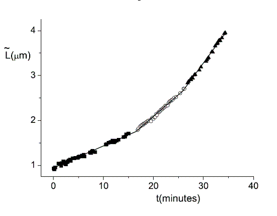
During the lifetime of a bacterium it elongates linearly in three distinct regimes.
We use single cell phase-contrast and fluorescence time-lapse microscopy to monitor morphological changes during the division of E. coli. To bypass the limitations of optical resolution, we process the images using pixel intensity values for edge detection. We study the dynamics of the constriction width, W, and find that its formation starts shortly after birth much earlier than can be detected by simply viewing phase-contrast images. A simple geometrical model is shown to reproduce the behavior of W(t). Moreover, the time-dependence of the cell length, L(t), consists of three linear regimes.